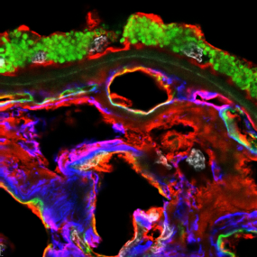
In a recent paper by Navratil et al, we examined the distribution and partial identities of sialic acids in human choroid, retinal pigment epithelium, photoreceptors, and lesions of age-related macular degeneration (AMD) using lectin histochemistry and microscopy.
Sialoglycoconjugates (glycoproteins or glycolipids capped by sialic acids) are known to play key roles in complement regulation, which is disrupted in AMD. Two forms of sialic acid, a-2,3 and a-2,6, have different distributions in the choroid, with both present on endothelial cell surfaces but only a-2,6 found in the intercapillary pillars surrounding the choriocapillaries. Both forms of sialic acid are found in foveal cones, but neither is present in cones outside the fovea. Basal laminar deposits (lesions that develop early in AMD) are consistently positive for both sialic acid forms. Partial identities of sialoglycoconjugates were also uncovered.
This study was published in Experimental Eye Research (https://doi.org/10.1016/j.exer.2025.110618).